Technical analysis, not a second launch story: Aleph Neuro has put one specific question in front of the brain-interface field with its June 24 post: can ultrasound localization microscopy, usually limited by the skull in adult humans, produce useful high-resolution brain images from outside the head?
Aleph does not name founders or executives in the material it published. What it does disclose is a company-level thesis. Aleph says the first limit on mind interfaces is not decoding software, but sensing hardware: fMRI can resolve brain activity but sits inside an MRI machine, implanted electrodes require surgery, and EEG or MEG can cover the head but produce lower-resolution signals. Aleph's bet is that neurovascular ultrasound can sit between those extremes by reading blood-flow changes tied to neural activity without opening the skull.
This explainer focuses on the mechanism behind the claim: microbubbles, localization microscopy, the 4-minute acquisition, the open-sourced reconstruction pipeline, and the still-unsolved problem of doing this repeatedly through human skulls. Aleph's public materials do not include a peer-reviewed Aleph paper, FDA clearance, a clinical validation package, or independent replication.
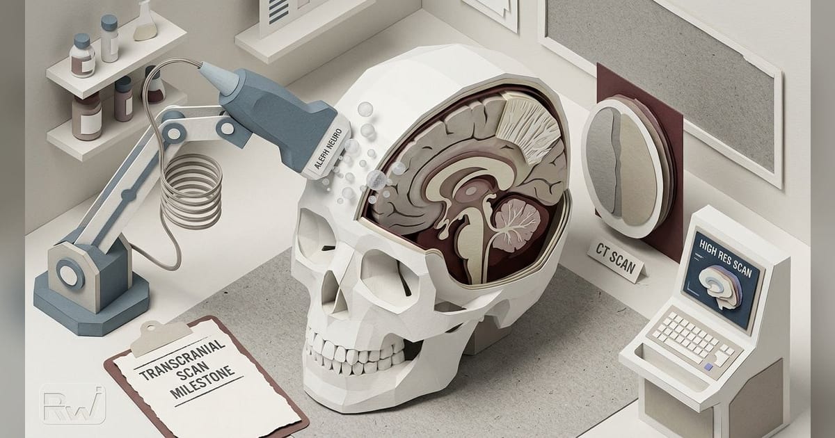
The image is vascular, not electrical
Aleph's image is not a direct recording of neurons firing. It is a vascular image. The premise is neurovascular coupling: when neurons become active, local blood flow and blood volume change. That is why Aleph compares its long-term direction to fMRI. Both approaches read a vascular proxy for neural activity rather than electrical spikes themselves.
That distinction matters. A high-detail vessel map can be a meaningful imaging milestone without proving that a system can decode thoughts, reconstruct visual scenes, or ship as a wearable brain-computer interface. Aleph frames its work against fMRI-based visual reconstruction systems such as MindEye, which reconstructed seen images from brain activity. But MindEye used fMRI. Aleph is arguing that the missing layer is portable, high-resolution sensing.
Aleph says neurovascular ultrasound could theoretically record "a million independent pixels throughout the brain, at less than a millimeter each." It also says even 1,000 invasive electrodes capture "at most 0.001% of the brain." Those are company claims, but they show the axis on which Aleph wants to compete: field of view plus resolution, not only electrode count or decoder performance.
Microbubbles are the signal amplifier
The result Aleph is showing is contrast-enhanced. In the post, Aleph says it infused microbubbles continuously during a 4-minute acquisition. The bubbles are sulfur hexafluoride pockets inside lipid shells. The National Cancer Institute describes sulfur hexafluoride lipid microspheres as contrast-enhancing agents that reflect ultrasound waves at the microsphere-tissue interface.
That reflection is the point. Red blood cells scatter ultrasound weakly. Microbubbles scatter strongly because the gas inside the lipid shell has acoustic properties very different from tissue. In raw ultrasound data, a bubble does not appear as a crisp dot. It appears as a blurred spot roughly set by the system's wavelength-scale resolution.
Ultrasound localization microscopy turns that defect into a measurement strategy. If bubbles are sparse enough that their blurred spots do not overlap too much, software can estimate the center of each spot with sub-pixel precision. As bubbles move through the vasculature, the system accumulates millions of localized bubble positions. Stack enough of those positions and the reconstructed vessel map can show structure below the usual diffraction limit.
Aleph says that is how it produced what it calls the world's first 3D image of ultrasound localization microscopy in a human brain through an intact skull, with "100 times greater volumetrically" resolution than comparable CT. The important caveat is that this is still Aleph's claim from its own technical post. The public record supplied with the announcement does not establish outside validation.
Localization is where the claim lives
The hardware matters, but Aleph's most inspectable claim sits in reconstruction. The company says it is open sourcing the pipeline and dataset behind the image. The GitHub repository, which redirects to alephneuro/microbubbles, provides code and sample data for ultrasound localization microscopy and instructions to reconstruct a 3D volume.
That is the credibility move. Outside researchers can inspect the processing choices behind the public image: how clutter is filtered, how bubbles are localized, how bubble positions are linked across frames, which tracks are retained, and how the 3D volume is rendered. Open code does not prove the biological or clinical claims, but it makes the technical artifact more auditable than a closed demo.
It also narrows the meaning of the milestone. Aleph is showing a contrast-enhanced structural vascular reconstruction, not a finished non-invasive brain interface. The difference is not semantic. A structural vascular image can be built by accumulating bubble positions over minutes. A brain-interface signal would need to capture functional changes with enough temporal resolution, repeatability, and signal quality to support decoding.
The skull is the hard part
Ultrasound is attractive because it can be cheaper and more compact than MRI and can reach deeper tissue than many optical techniques. The skull is why adult human brain ultrasound remains difficult.
Research on transcranial ultrasound has long shown that the skull attenuates and distorts ultrasound waves. A study of acoustic properties across intact human skulls found that ultrasound transmission and phase distortion vary substantially across regions and individuals. That means a system has to handle not just one skull, but differences across people, bone thicknesses, and acoustic windows. (Acoustic properties across the human skull)
Aleph acknowledges the boundary indirectly. The company says neurovascular ultrasound has produced strong results when the skull is removed, but that the challenge is doing it with the skull intact. Prior work explains why the modality keeps drawing attention. A 2021 Neuron paper, "Single-trial decoding of movement intentions using functional ultrasound neuroimaging", reported movement-intention decoding using functional ultrasound. Aleph's own post also points to later work showing neurovascular ultrasound results when the skull is removed. Aleph is trying to move that promise toward adult human transcranial imaging.
The skull problem is not only loss of signal. It is also distortion. Ultrasound waves can arrive with phase errors after passing through bone, which can blur or misplace reconstructed features unless corrected. For localization microscopy, that matters because the final image depends on estimating bubble centers accurately across many frames.
Why contrast-free imaging is the real target
Aleph says the microbubble result is a step toward contrast-free neurovascular imaging. That target would remove injected bubbles from the workflow and instead extract signal from red blood cells moving through vessels.
The tradeoff is signal strength. Aleph says red blood cells scatter far less than microbubbles, which makes contrast-free imaging harder. Its proposed answer is not only better probes, but better use of raw data. Aleph says a standard ultrasound probe receives terabytes of data per hour, while typical processing pipelines compress that stream down to about 0.1% of the original. The company argues that end-to-end machine learning trained on large datasets can recover signal that hand-engineered pipelines discard.
That thesis is plausible in the same broad way early computer-vision arguments were plausible: sensors collect more information than legacy algorithms use, and learned systems may extract more structure from the stream. But the burden of proof is high. Aleph would have to show that the recovered signal is real physiology, not reconstruction artifact; that it generalizes across subjects; and that it can track functional changes at useful speeds.
What would validate it
Aleph's technical claim has three separable tests.
First, reproducibility through intact adult skulls. One high-detail image can show feasibility. A platform has to work across skull geometries, acoustic windows, ages, and subjects. The question is not whether a best-case scan can be reconstructed, but whether the method survives ordinary biological variation.
Second, functional relevance. Microbubble localization can produce detailed vascular maps, but a brain interface needs changes over time. Aleph's stated destination depends on contrast-free neurovascular imaging that can extract weaker red-blood-cell signal with enough temporal and spatial resolution to support decoding.
Third, device translation. The public post does not establish a commercial product, clinical workflow, regulatory path, or customer deployment. It shows a research pipeline and a dataset. Turning that into a safe, repeatable device outside a controlled acquisition setup is a separate engineering and regulatory problem.
Aleph has not disclosed funding, investors, valuation, headquarters, headcount, leadership, product status, or customer deployments in the materials available. It has disclosed a dataset, code, a 4-minute microbubble acquisition, and a specific through-skull reconstruction claim. For now, that is the useful way to read the announcement: not as proof of mind reading, but as a testable attempt to push ultrasound localization microscopy through the adult human skull.