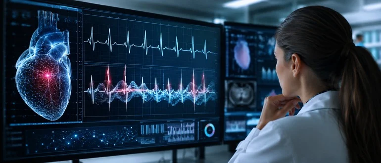
TL;DR — Key Takeaways
- AI is extracting hidden diagnostic and predictive signals from routine ECGs that cardiologists may not be able to detect visually.
- Cardiovolt, developed from research at Imperial College London, was trained on millions of ECGs linked to patient histories and outcomes.
- The technology reports high accuracy for detecting conditions including left ventricular dysfunction, aortic stenosis and tricuspid regurgitation.
About 1 billion electrocardiograms (ECGs) are performed around the world each year, each one capturing an electrical portrait of the human heart. ECGs also contain hidden patterns, and it is those patterns that could give cardiologists an advantage in preventing cardiovascular diseases.
A new AI tool, developed by researchers at Imperial College London’s National Heart and Lung Institute and trained on millions of ECGs, can spot signs of heart disease from an ECG in about two seconds.
“Every ECG contains far more information than conventional interpretation reveals,” states Cardiovolt.ai, a company created to promote the new AI tool. “Cardiovolt extracts subtle digital biomarkers from existing recordings, no additional hardware, integrating seamlessly into existing clinical workflows.”
Dr. Arunashis Sau, an academic clinical lecturer at the institute and the chief scientific officer of Cardiovolt.ai, called the AI tool superhuman, “doing things that no cardiologist, no matter how expert, can do.”
Cardiovolt was trained on millions of ECGs linked to patients’ medical histories. By comparing electrical patterns with diagnoses and long-term outcomes, researchers trained the model to uncover particular diseases and future health risks.
A cardiologist can scrutinize an individual ECG, but no human can realistically compare millions of recordings with years of medical outcomes and search for minute variations across them. Cardiovolt can perform that analysis repeatedly and at enormous scale, identifying patterns that might otherwise remain buried in the data.
Cardiovolt reports diagnostic accuracy of 93% for left ventricular dysfunction, 82% for aortic stenosis and 81% for tricuspid regurgitation. Its predictive models report 86% accuracy for complete heart block and 82% for both mortality risk and atrial fibrillation.
Cardiovolt says the technology is still in the regulatory process and is not yet ready for routine clinical use. Its intended role is to identify patients whose ECGs contain signals warranting further investigation, potentially directing them to more definitive testing before a disease becomes clinically obvious.
“If someone comes in to have an ECG, we want to pick up underlying heart failure or valve disease that would never be picked up by a human doctor,” said Professor Fu Siong Ng, a cardiologist at Imperial College London and Cardiovolt’s chief medical officer. If the AI identifies a possible problem, he said, an echocardiogram can then be used to confirm it.
An echocardiogram uses ultrasound to create images of the heart and can reveal structural abnormalities that an ECG cannot directly show. If AI can identify which patients are most likely to have those abnormalities, a routine ECG could become a much more powerful screening tool.
“A 10-second ECG, once a blunt screening tool, may soon become one of the most informative tests in medicine,” Professor Ng said.
Cardiovolt is part of a much larger movement in medicine in which AI is being used to detect patterns that are difficult for humans to perceive or process at scale. Researchers are applying similar approaches to medical images, pathology slides, genomic data and electronic health records, where the volume and complexity of information can overwhelm even highly trained specialists.
One recent study suggests that the same principle could extend to an entirely different kind of medical image: the mammogram.
Researchers in Israel used AI to analyze 97,364 mammograms from 29,921 women with an average age of 54, looking for patterns associated with cardiovascular disease. The machine-learning model identified women with a history of stroke 86% of the time. It also distinguished women with high blood pressure and coronary heart disease from those without the conditions with reported accuracies of 79% and 78%, respectively. The results were consistent regardless of the women’s age or whether they also had cancer.
The findings raise the possibility that a test already used to screen for breast cancer could someday also flag women at elevated risk of cardiovascular disease. Because mammograms are already performed on hundreds of millions of women, researchers say the approach could potentially provide an additional source of cardiovascular information without requiring another imaging examination.
The researchers are still working to improve the model’s accuracy and reduce false results, meaning the technology is not ready to be used as a stand-alone clinical test. But the study illustrates a growing theme in medical AI: The information needed to identify disease may already exist inside tests doctors routinely perform. The challenge is teaching machines to recognize it.